
Explore the stretch reflex, including its definition, physiological mechanism, reflex arc, functions, clinical applications in physiotherapy and factors affecting the reflex response.
Definition
The stretch reflex is also known as the myotatic reflex. It is a monosynaptic spinal reflex because the primary sensory neuron forms a direct excitatory synapse with the alpha motor neuron supplying the stretched muscle. The stretch reflex is an involuntary, automatic contraction of a muscle in response to its sudden stretching. It helps maintain muscle tone, regulate muscle length, and stabilize posture.
Receptors Involved
Muscle spindle: Detects muscle length and changes in muscle length.
Group Ia sensory afferent fibres: Primarily convey information about the dynamic component of stretch, including the rate of muscle lengthening.
Group II sensory afferent fibres: Primarily convey information about sustained muscle length.
Alpha motor neurons: Supply extrafusal muscle fibres and produce muscle contraction.
Mechanism of the Stretch Reflex
The stretch reflex sequence:
- Stretch stimulus: A muscle is suddenly stretched.
- Activation of muscle spindles: The muscle spindle detects the change in muscle length.
- Sensory impulse transmission: Group Ia sensory fibres carry impulses to the spinal cord through the dorsal root.
- Synaptic transmission: The Ia sensory neuron forms an excitatory synapse directly with an alpha motor neuron in the spinal cord.
- Motor impulse transmission: The alpha motor neuron carries impulses through the ventral root to the same muscle.
- Muscle contraction: The stretched muscle contracts, opposing the initial stretch.
- Reciprocal inhibition: Through an inhibitory interneuron, the antagonist muscle is inhibited, facilitating the movement produced by the agonist.
- The direct sensory-to-motor pathway produces the monosynaptic component of the reflex, whereas reciprocal inhibition involves an additional interneuron.
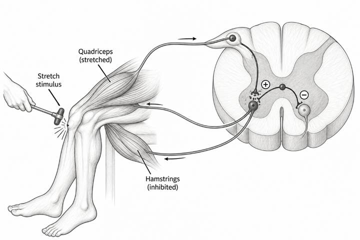
Pathway of the Stretch Reflex
Stretch stimulus → Muscle spindle activation → Ia sensory afferent → Spinal cord → Alpha motor neuron → Neuromuscular junction → Contraction of the stretched muscle.
At the same time, inhibitory interneurons help reduce activity in the antagonist muscle.
Types of Stretch Reflex
Phasic Stretch Reflex: The phasic stretch reflex is a rapid, short-lasting muscle contraction in response to a sudden stretch. Eg: The knee-jerk reflex following a tap on the patellar tendon.
Tonic Stretch Reflex: The tonic stretch reflex refers to the sustained reflex activity associated with maintaining muscle length and muscle tone during prolonged or continuous stretch. Eg: Reflex contributions to the activity of postural muscles during standing.
Clinical Examples: Knee-Jerk Reflex, Ankle-Jerk Reflex
Functions of the Stretch Reflex
Maintenance of muscle tone: Helps maintain a baseline level of muscle activity.
Postural control: Contributes to the maintenance of upright posture.
Regulation of muscle length: Opposes sudden changes in muscle length.
Joint stability: Helps stabilize joints during unexpected disturbances.
Movement control: Contributes to the coordination of muscle activity during functional movements.
Protection against sudden lengthening: Helps resist rapid muscle stretch, although it is not an absolute protective mechanism.
Factors Affecting the Stretch Reflex
Rate of stretch: A rapid stretch generally produces a stronger dynamic response than a slow stretch of comparable magnitude.
Magnitude of stretch: Greater stretch may increase spindle activation, within physiological limits.
Muscle spindle sensitivity: Influenced by gamma motor neuron activity.
Descending control: Signals from higher centres modify spinal reflex excitability.
Muscle fatigue and physiological state: May alter the response.
Neurological conditions: Upper motor neuron lesions may increase tendon reflexes, whereas lesions affecting the reflex arc may reduce or abolish them.
Age and individual variation: Reflex responses can vary among individuals and across the lifespan.
Clinical Significance in Physiotherapy
- The stretch reflex is important in neurological examination and rehabilitation.
- Deep tendon reflex testing: Helps assess the integrity of the reflex arc and relevant spinal segments.
- Assessment of upper motor neuron lesions: Exaggerated tendon reflexes may occur in conditions involving reduced descending inhibition.
- Assessment of lower motor neuron lesions: Reduced or absent reflexes may occur when the sensory or motor components of the reflex arc are damaged.
- Spasticity assessment: Increased stretch-reflex excitability contributes to the velocity-dependent resistance to passive movement observed in spasticity.
- Rehabilitation planning: Understanding reflex activity helps physiotherapists select appropriate positioning, movement strategies, and interventions for patients with altered muscle tone.
References
- Kisner, C., Colby, L. A., & Borstad, J. (2017). Therapeutic Exercise: Foundations and Techniques (7th ed.). F. A. Davis Company.
- Dutton, M. (2020). Dutton’s Orthopaedic Examination, Evaluation, and Intervention (5th ed.). McGraw Hill.
- Neumann, D. A. (2017). Kinesiology of the Musculoskeletal System: Foundations for Rehabilitation (3rd ed.). Elsevier.
For more notes click on: www.physiotherapyphd.com

