
Explore pectoralis major stretching techniques, including positioning, step-by-step instructions, stretching duration, precautions and benefits for chest flexibility, shoulder mobility and posture.
Definition
Pectoralis major stretching is a therapeutic technique used to lengthen the pectoralis major muscle and improve flexibility of the anterior chest wall. It helps reduce muscle tightness, improve shoulder mobility, and facilitate an upright posture.
Anatomy of the Pectoralis Major
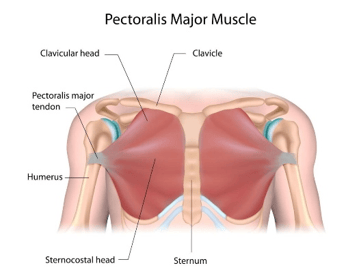
The pectoralis major is a large, fan-shaped muscle located on the anterior aspect of the chest.
Origin: Medial half of the clavicle, anterior surface of the sternum, costal cartilages of the upper six ribs (commonly described as ribs 1–6), and aponeurosis of the external oblique muscle.
Insertion: Lateral lip of the intertubercular sulcus of the humerus.
Nerve supply: Lateral and medial pectoral nerves (C5–T1).
Actions:
- Shoulder horizontal adduction.
- Shoulder adduction.
- Medial rotation of the shoulder.
- Clavicular head assists in shoulder flexion.
- Sternocostal head assists in shoulder extension from a flexed position.
Pectoralis Major Stretching- uses
- To increase the flexibility of the pectoralis major muscle.
- To improve shoulder range of motion, particularly horizontal abduction.
- To reduce tightness of the anterior chest muscles.
- To facilitate improved shoulder and scapular positioning.
- To assist in correcting rounded shoulder posture when associated with pectoral muscle tightness.
Indications
- Tightness of the pectoralis major muscle.
- Rounded shoulder posture associated with anterior chest tightness.
- Restricted shoulder horizontal abduction.
- Reduced flexibility following prolonged sitting or desk work.
- Muscle tightness associated with repetitive activities involving forward shoulder positioning.
- Rehabilitation programs requiring restoration of shoulder mobility, when permitted by the treating clinician.
Contraindications
- Acute pectoralis major muscle strain or tear.
- Recent pectoral or shoulder surgery without clearance.
- Acute shoulder injury with significant pain or instability.
- Suspected fracture or acute soft-tissue injury.
Precautions
- Avoid forceful stretching and sudden movements.
- Do not stretch through sharp pain or pinching in the shoulder.
- Use a smaller range in patients with shoulder instability.
- Following breast, chest wall, or shoulder surgery, follow the prescribed rehabilitation protocol.
- Stop if numbness, tingling, or unusual symptoms develop in the arm.
Self-Stretching Techniques of Pectoralis Major Stretching
A. Doorway Stretch
Starting position: Standing in a doorway.
Procedure:
- Ask the patient to stand in a doorway with the feet comfortably apart.
- Place the forearm against the doorframe, keeping the elbow approximately at shoulder height and bent to 90 degrees.
- Position the elbow and forearm securely against the frame.
- Ask the patient to take a small step forward until a gentle stretch is felt across the front of the chest.
- Keep the trunk upright and avoid excessive arching of the lower back.
- Maintain the stretch for 20–30 seconds.
- Return slowly to the starting position.
- Repeat 2–3 times on each side, as tolerated.
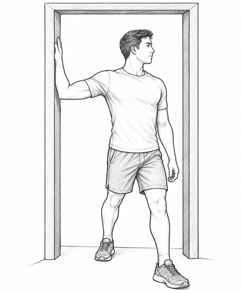
Common errors:
- Leaning too far forward.
- Elevating the shoulder toward the ear.
- Arching the lower back excessively.
- Stretching until pain occurs.
B. Corner Stretch
Starting position: Standing facing a corner of a room.
Procedure:
- Ask the patient to stand facing the corner.
- Place both forearms on the adjoining walls, with the elbows approximately at shoulder level.
- Position the feet in a comfortable staggered stance.
- Gently lean the body forward toward the corner.
- Keep the head and trunk aligned and the shoulders relaxed.
- Continue until a mild stretch is felt across the anterior chest.
- Hold for 20–30 seconds.
- Return to the starting position and repeat 2–3 times.
Target muscle: Bilateral pectoralis major, with contribution from other anterior shoulder tissues.
Precaution: Avoid excessive forward leaning or forcing the shoulders into an uncomfortable position.
C. Supine Pectoral Stretch
Starting position: Lying supine on a treatment table.
Procedure:
- Ask the patient to lie on their back with the head and trunk supported.
- Position the arm out to the side at a comfortable angle.
- Keep the elbow slightly bent or positioned as instructed.
- Allow the arm to move gently toward horizontal abduction, within the available pain-free range.
- Maintain a relaxed shoulder and avoid excessive arching of the trunk.
- Hold for 20–30 seconds.
- Return the arm slowly to the starting position.
- Repeat 2–3 times, as tolerated.
Target muscle: Pectoralis major, depending on the degree of shoulder abduction and rotation.
Precaution: Do not force the arm below the level of the treatment table if this causes pain or shoulder instability.
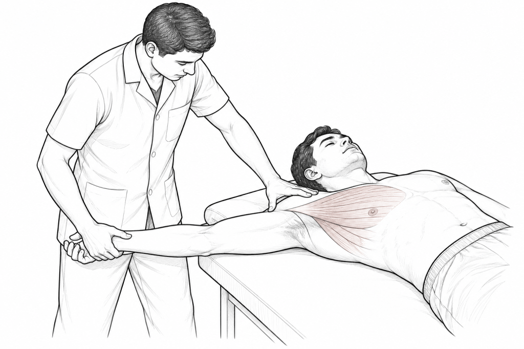
D. Manual Pectoralis Major Stretch
Starting position: Patient lying supine on a treatment table.
Procedure:
- Ask the patient to lie comfortably in the supine position.
- Stabilize the thorax and scapular region as appropriate.
- Support the patient’s arm and position the shoulder in a comfortable degree of abduction and external rotation.
- Gently move the arm toward horizontal abduction.
- Apply a slow, controlled stretch without producing pain.
- Hold for 20–30 seconds, according to the patient’s tolerance and clinical condition.
- Release gradually and return the arm to the resting position.
- Repeat as indicated.
Precaution: This technique should be performed by a trained physiotherapist, particularly in patients with shoulder pathology or a history of surgery.
Dosage and Application
| Parameter | General recommendation |
| Duration | 20–30 seconds per repetition |
| Repetitions | 2–3 per side or position |
| Intensity | Mild, comfortable stretching sensation |
| Frequency | Once or twice daily, depending on need and tolerance |
| Technique | Slow, controlled, static stretching |
| Breathing | Normal breathing without holding the breath |
The dosage should be modified according to the patient’s condition, pain response, and rehabilitation goals.
Mechanism of Stretching
Pectoralis major stretching applies a controlled tensile force to the muscle–tendon unit. With appropriate positioning and sustained stretching, it may improve stretch tolerance and contribute to increased muscle flexibility and shoulder mobility.
The response depends on the stretching intensity, duration, frequency, and the individual’s clinical condition. Stretching alone does not guarantee correction of rounded shoulder posture; strengthening of the scapular retractors and other postural muscles may also be required.
Assessment of Pectoralis Major Tightness
Pectoralis major tightness can be assessed through observation and clinical examination.
Common assessment approach:
- Position the patient in supine lying.
- Observe the resting position of the shoulder and arm.
- Assess whether the shoulder rests in an elevated or forward position.
- Passively move the arm toward horizontal abduction, within a safe range.
- Observe the available movement, compensatory trunk movement, and symptoms.
- Compare both sides and document the findings.
Interpretation: Restricted movement, a reproducible feeling of anterior chest tightness, or compensatory movement may suggest reduced flexibility. However, these findings are not specific to pectoralis major tightness and should be interpreted alongside the patient’s history and a full shoulder examination.
Patient Education and Home Program
- Perform stretching slowly and consistently.
- Maintain a comfortable posture throughout the exercise.
- Avoid bouncing or jerking movements.
- Stop if sharp pain or unusual neurological symptoms occur.
- Combine stretching with appropriate strengthening and postural exercises when indicated.
Seek professional assessment if pain or restricted movement persists.
References
1. Kisner C, Colby LA, Borstad J. Therapeutic Exercise: Foundations and Techniques. 7th ed. Philadelphia: F.A. Davis Company; 2017. Chapter 4: Stretching for Improved Mobility.
2. American College of Sports Medicine. ACSM’s Guidelines for Exercise Testing and Prescription. 8th ed. Philadelphia: Lippincott Williams & Wilkins; 2010.
For more pdf/notes click on: www.physiotherapyphd.com

